- About
- Specialists
-
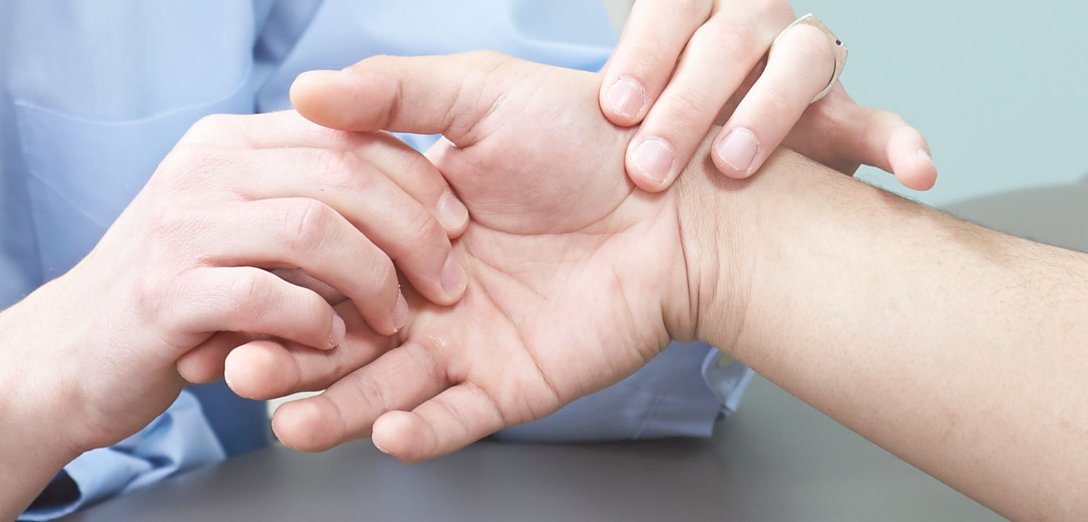
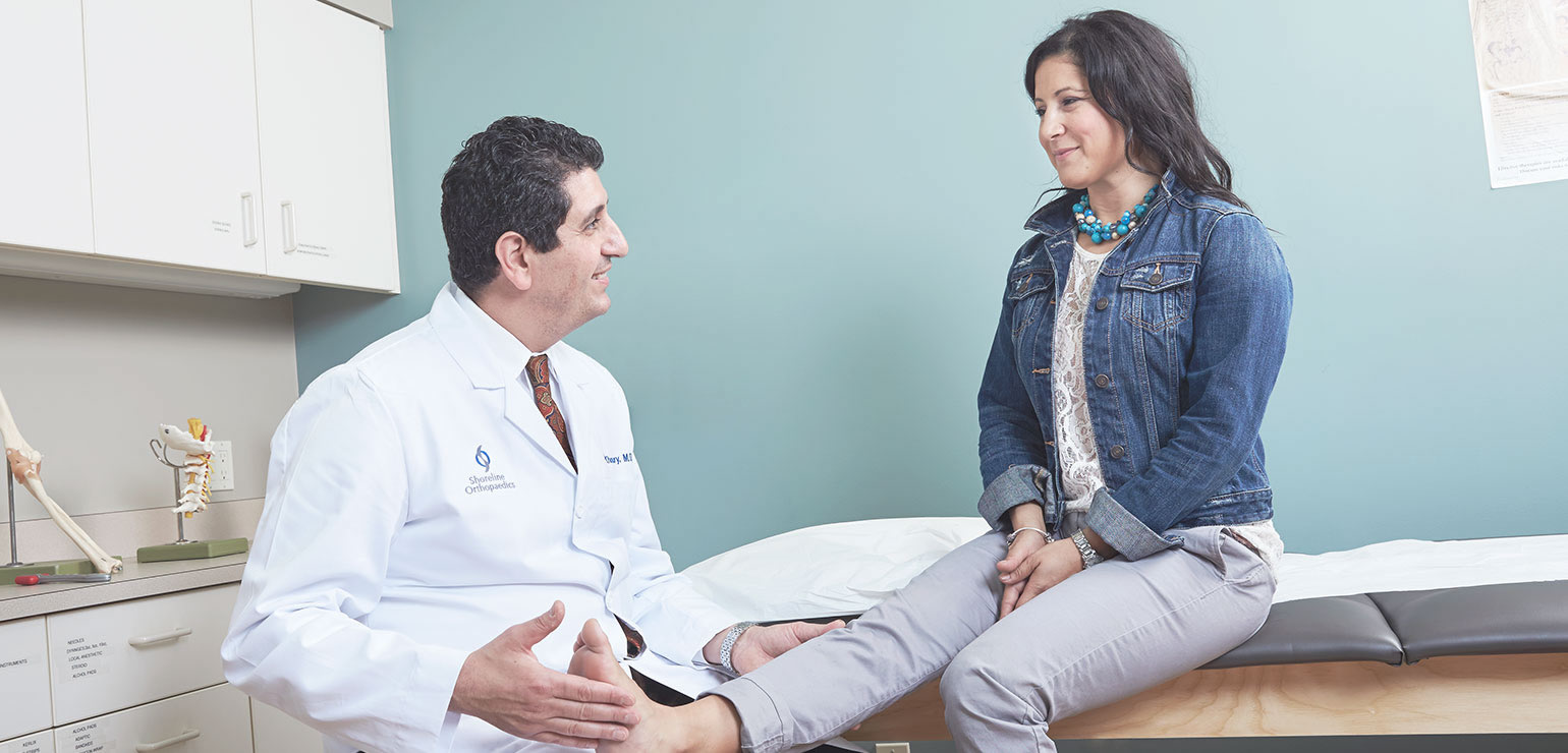
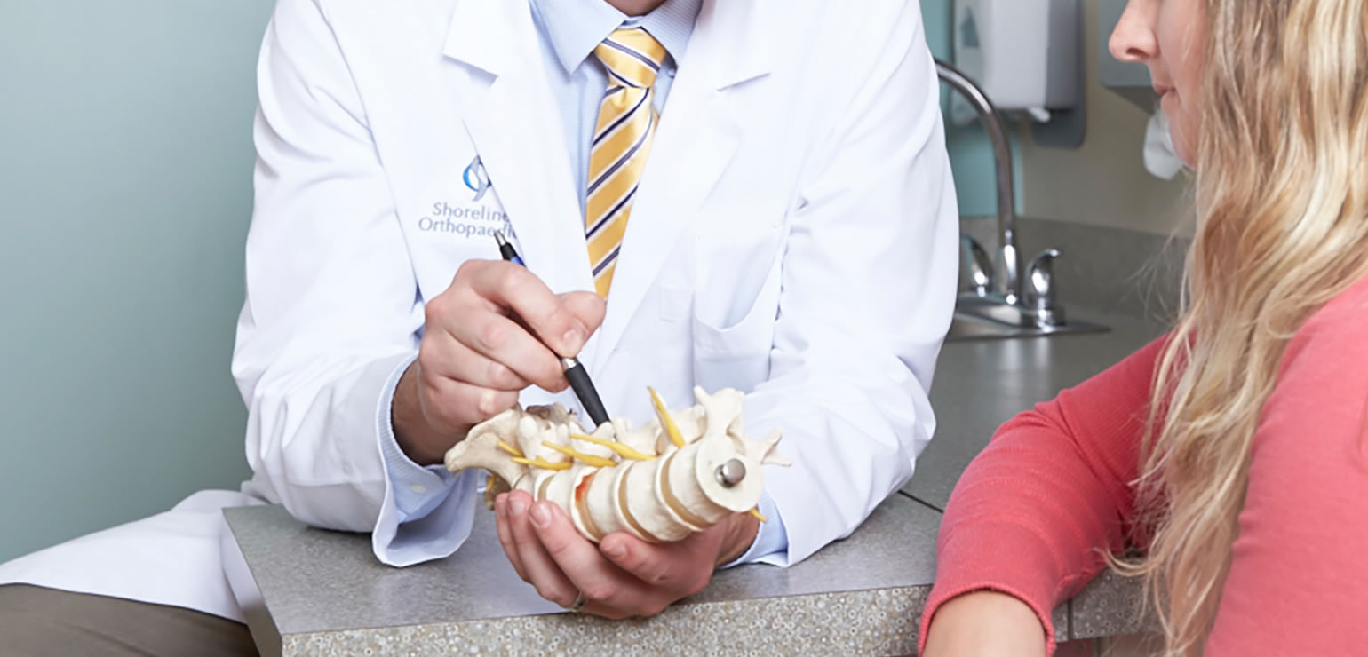
At Shoreline Orthopaedics, our orthopaedic surgeons use a truly collaborative approach so our patients have the benefit of multiple expert opinions, without having to go elsewhere to obtain them.
-
- Specialties
- Arthritis
- Diagnostics
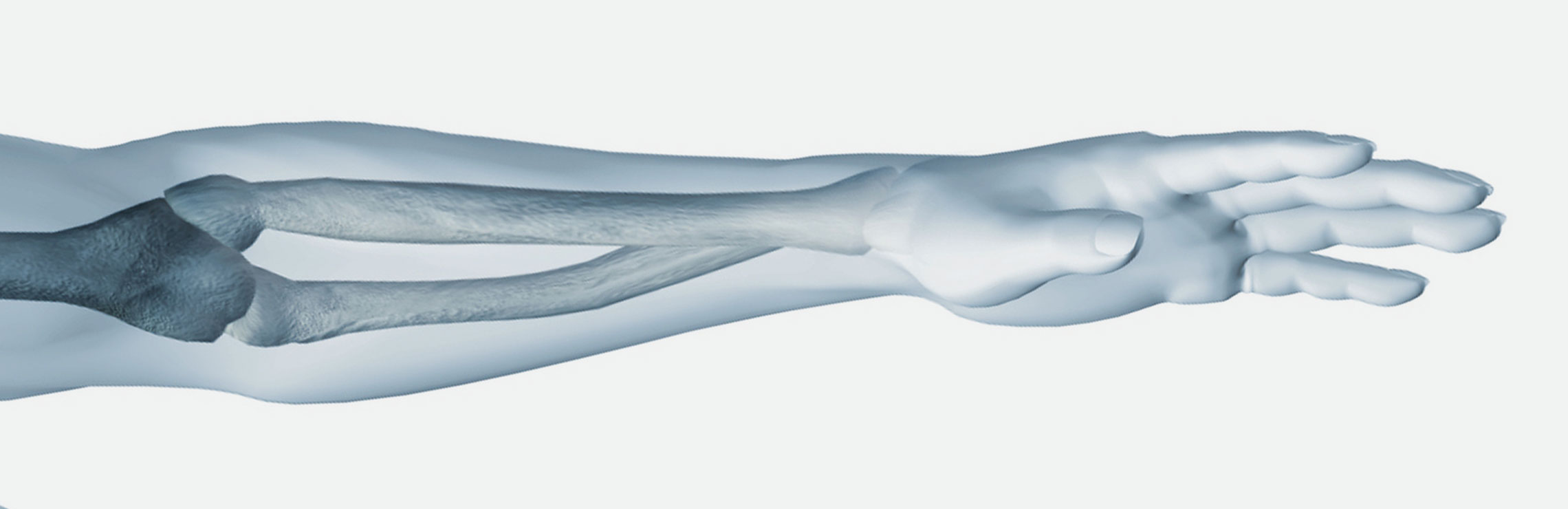
- Elbow

- Biceps Tendon Tear at the Elbow
- Elbow Arthroscopy
- Elbow (Olecranon) Bursitis
- Golfer's Elbow (Medial Epicondylitis)
- Loose Body in the Elbow
- Radial Head Fractures of the Elbow
- Tennis Elbow (Lateral Epicondylitis)
- Throwing Injuries to the Elbow in Children
- Ulnar Nerve Entrapment at the Elbow (Cubital Tunnel Syndrome)
- Foot & Ankle
- Achilles Tendinitis
- Achilles Tendon Rupture
- Ankle Arthritis
- Ankle Sprain
- Bone Health
- Bunions
- Cavovarus Foot Deformity
- Equinus
- Hallux Rigidus (Stiff Big Toe)
- Morton's Neuroma
- Peroneal Tendon Injuries
- Pes Plano Valgus (Flexible Flatfoot in Children)
- Plantar Fasciitis
- Posterior Tibial Tendon Dysfunction
- Sever's Disease
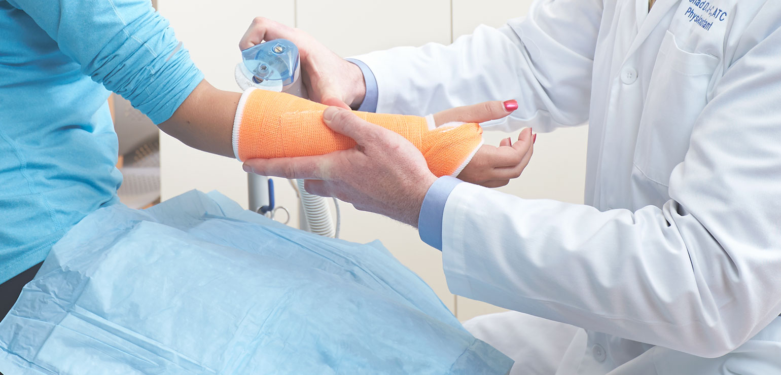
- Fractures, Sprains and Strains
- AC Joint Issues
- Ankle Sprain
- Bone Health
- Cervical Fracture (Broken Neck)
- Finger Fracture
- Fractures
- Fracture of the Thoracic and Lumbar Spine
- Growth Plate Fractures
- Hand Fracture
- Low Back Pain
- Lumbar Back Strain
- Neck Sprains and Strains
- Osteoporosis and Spinal Fractures
- Radial Head Fractures of the Elbow
- Shoulder Separation
- Sprains and Strains
- Stress Fracture
- Thumb Fracture
- Thumb Sprain
- Wrist, Distal Radius Fracture
- Wrist, Scaphoid Fracture
- Wrist Sprains
- Hand and Wrist

- Bone Health
- Carpal Tunnel Syndrome
- Dupuytren's Contracture
- Extensor Tendon Lacerations
- Hand and Wrist Arthritis
- Hand and Wrist Tendinitis
- Finger Fracture
- Hand Fracture
- Ganglion Cyst
- Thumb Fracture
- Wrist, Distal Radius Fracture
- Wrist, Scaphoid Fracture
- De Quervain's Tendinitis
- Flexor Tendon Injuries
- Nerve Injuries
- Thumb Sprain
- Trigger Finger
- Wrist Arthroscopy
- Wrist Sprains
- Hip
- Joint Replacement and Revision
- Joint Disorders
- AC Joint Inflammation
- AC Joint Issues
- Ankle Arthritis
- Anterior or Posterior Hip Replacement
- Arthritis
- Articular Cartilage Restoration
- Biceps Tendinitis
- Biceps Tendon Tear at the Elbow
- Bunions
- Elbow Arthroscopy
- Elbow (Olecranon) Bursitis
- Femoral Acetabular Impingement (FAI) & Labral Tear (Hip)
- Frozen Shoulder (Adhesive Capsulitis)
- Ganglion Cyst
- Golfer's Elbow (Medial Epicondylitis)
- Hallux Rigidus (Stiff Big Toe)
- Hand and Wrist Arthritis
- Hip Bursitis
- Hip Arthroscopy
- Hip Osteonecrosis
- Hip Resurfacing
- Knee Arthroscopy
- Knee Osteotomy
- Knee Osteonecrosis
- Loose Body in the Elbow
- Osteochondritis Dissecans
- Partial Knee Replacement
- Patellofemoral Pain Syndrome
- Radial Head Fractures of the Elbow
- Shoulder Arthroscopy
- Shoulder Dislocation
- Shoulder Impingement
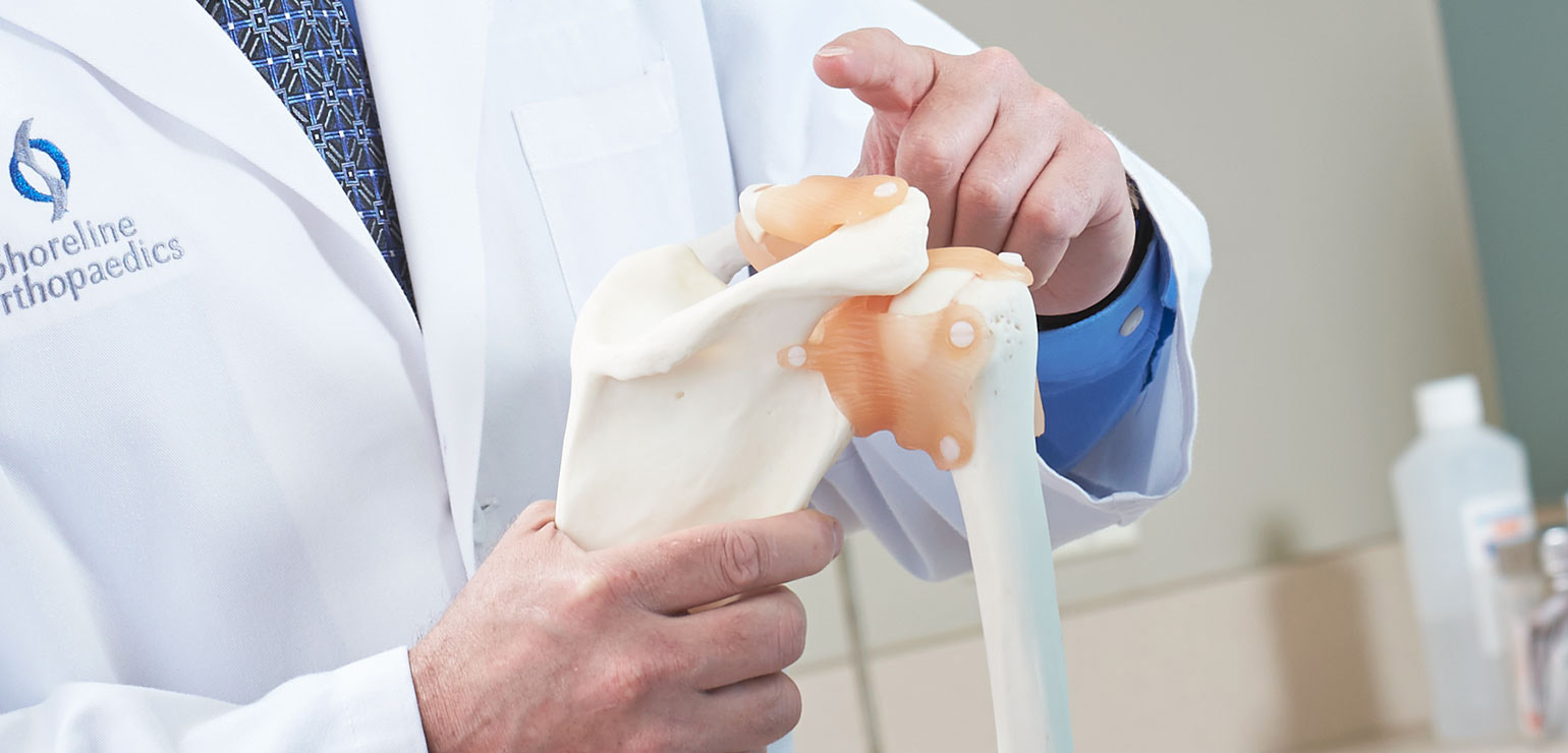
- Shoulder Replacement
- Shoulder Separation
- SLAP Tear
- Tennis Elbow (Lateral Epicondylitis)
- Total Hip Replacement
- Total Knee Replacement
- Ulnar Nerve Entrapment at the Elbow (Cubital Tunnel Syndrome)
- Unstable Kneecap (Patella Instability) Procedures
- Wrist Arthroscopy
- Ligament Disorders
- Muscle Disorders
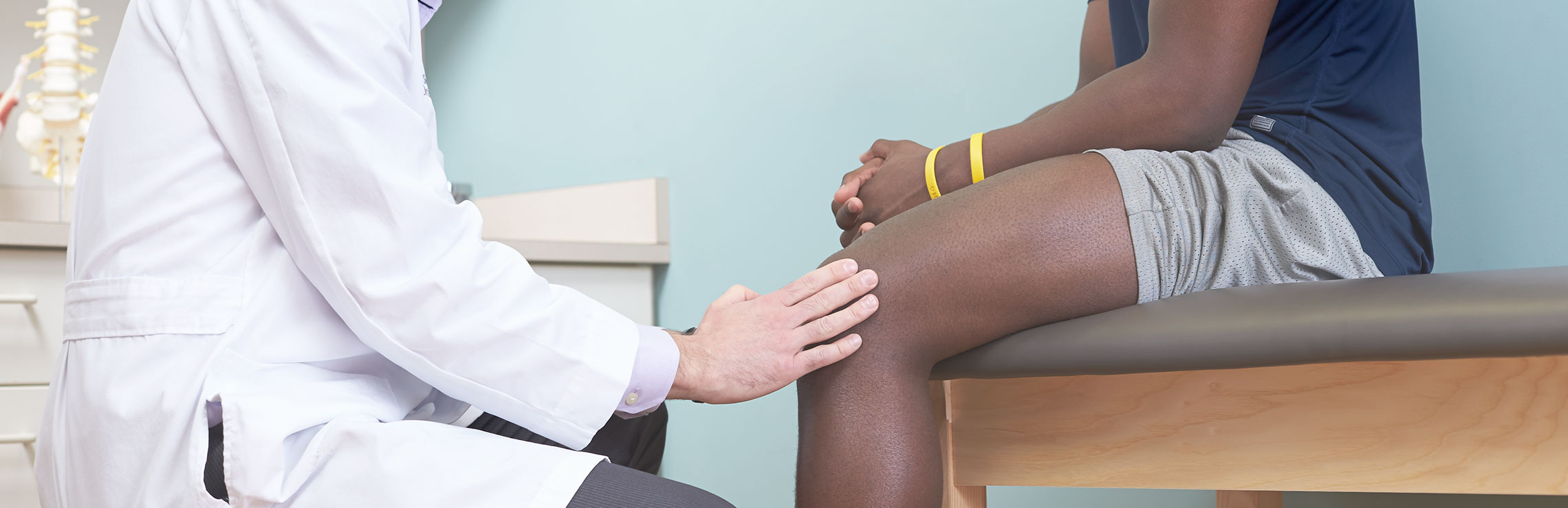
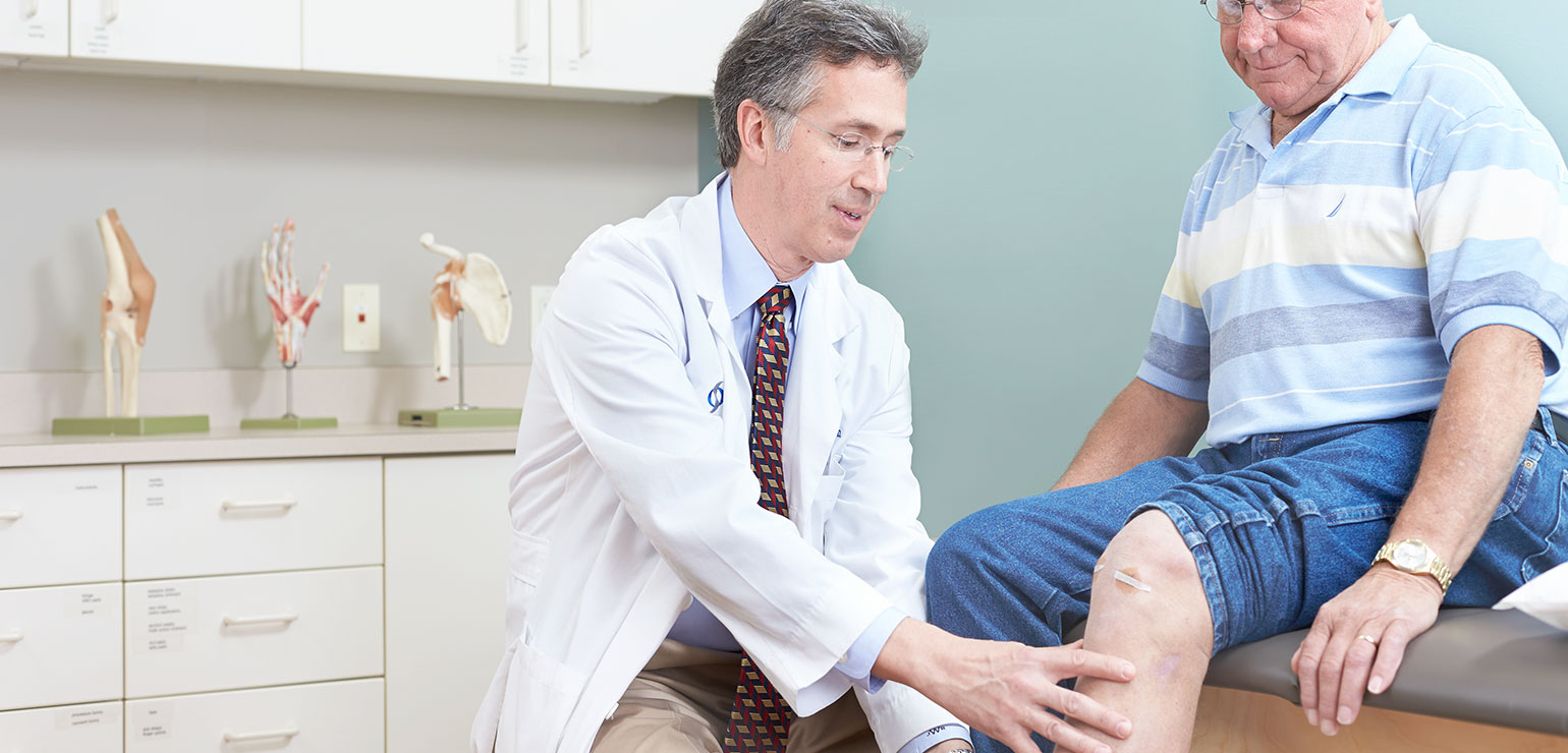
- Knee
- ACL (Anterior Cruciate Ligament) Injuries and Reconstruction
- Articular Cartilage Restoration
- Collateral Ligament Injuries (MCL, LCL)
- Combined Knee Ligament Injuries
- Jumper's Knee
- Knee Arthritis
- Knee Arthroscopy
- Meniscal Tears
- Osgood-Schlatter Disease
- Osteochondritis Dissecans
- Knee Osteonecrosis
- Knee Osteotomy
- Partial Knee Replacement
- Patella Tendinitis and Patella Tendinosis
- Patellofemoral Pain Syndrome
- PCL (Posterior Cruciate Ligament) Injuries and Reconstruction
- Total Knee Replacement
- Unstable Kneecap (Patella Instability) Procedures
- Minimally Invasive Surgery (Arthroscopy)
- Neck and Back (Spine)
- Backpack Safety
- Bone Health
- Cervical Fracture (Broken Neck)
- Cervical Radiculopathy (Pinched Nerve)
- Cervical Spondylosis (Arthritis of the Neck)
- Cervical Spondylotic Myelopathy (Spinal Cord Compression)
- Fracture of the Thoracic and Lumbar Spine
- Herniated Disk
- Kyphosis (Roundback) of the Spine
- Low Back Pain
- Lumbar Back Strain
- Lumbar Spinal Stenosis
- Neck Sprains and Strains
- Osteoporosis and Spinal Fractures
- Preventing Back Pain
- Sacroiliac Joint Dysfunction (SI Joint Pain)
- Sciatica
- Scoliosis
- Spondylolysis and Spondylolisthesis
- Pediatric Injuries
- Backpack Safety
- Bone Joint and Muscle Infections in Children
- Golfer's Elbow
- Growth Plate Fractures
- High School Sports Injuries
- Jumper's Knee
- Kyphosis (Roundback) of the Spine
- Osgood-Schlatter Disease
- Osteochondritis Dissecans
- Overuse Injuries in Children
- Patella Tendinitis and Patella Tendinosis
- Pes Plano Valgus (Flexible Flatfoot in Children)
- Scoliosis
- Sever's Disease
- Spondylolysis and Spondylolisthesis
- Throwing Injuries to the Elbow in Children
- Physical Medicine and Rehabilitation
- PM&R or Physiatry Overview
- AC Joint Issues
- Arthritis Overview
- Biceps Tendinitis
- Carpal Tunnel Syndrome
- Cervical Radiculopathy (Pinched Nerve)
- Cervical Spondylosis (Arthritis of the Neck)
- Cervical Spondylotic Myelopathy
- Femoral Acetabular Impingement (FAI) & Labral Tear of the Hip
- Fibromyalgia
- Herniated Disc
- Hip Arthritis
- Hip Bursitis
- Knee Arthritis
- Kyphosis (Roundback) of the Spine
- Neck Sprains & Strains
- Osteoporosis & Spinal Fractures
- Patellofemoral Pain Syndrome (Runner's Knee)
- Sacroiliac Joint Dysfunction (SI Joint Pain)
- Sciatica
- Scoliosis
- Shoulder Arthritis
- Shoulder Impingement
- Spondylolysis & Spondylolisthesis
- Shoulder
- Sports Medicine (General)
- Sports Medicine Overview
- Contusions or Bruises
- Cramps or Charley Horse
- Fractures
- Growth Plate Fractures
- Hamstring Injuries
- High School Sports Injuries
- Osteochondritis Dissecans
- Overuse Injuries in Children
- Platelet-Rich Plasma (PRP)
- Posterior Tibial Tendon Dysfunction
- Sprains and Strains
- Stress Fracture
- Throwing Injuries to the Elbow in Children
- Sports Medicine (Upper Body)
- AC Joint Inflammation
- AC Joint Issues
- Biceps Tendinitis
- Biceps Tendon Tear at the Elbow
- Cervical Fracture (Broken Neck)
- Golfer's Elbow (Medial Epicondylitis)
- Loose Body in the Elbow
- Rotator Cuff Tear
- Shoulder Dislocation
- Shoulder Impingement
- Shoulder Separation (AC Joint Sprain)
- SLAP Tear
- Tennis Elbow (Lateral Epicondylitis)
- Throwing Injuries to the Elbow in Children
- Thumb Sprain
- Wrist Sprains
- Sports Medicine (Lower Body)
- Achilles Tendinitis
- Achilles Tendon Rupture
- ACL (Anterior Cruciate Ligament) Injuries and Reconstruction
- Ankle Sprain
- Collateral Ligament Injuries (MCL, LCL)
- Combined Knee Ligament Injuries
- Femoral Acetabular Impingement (FAI) & Labral Tear of the Hip
- Jumper's Knee
- Osgood-Schlatter Disease
- Patella Tendinitis and Patella Tendinosis
- Patellofemoral Pain Syndrome
- PCL (Posterior Cruciate Ligament) Injuries and Reconstruction
- Peroneal Tendon Injuries
- Posterior Tibial Tendon Dysfunction
- Sever's Disease
- Spondylolysis and Spondylolisthesis
- Thigh Muscle Strain

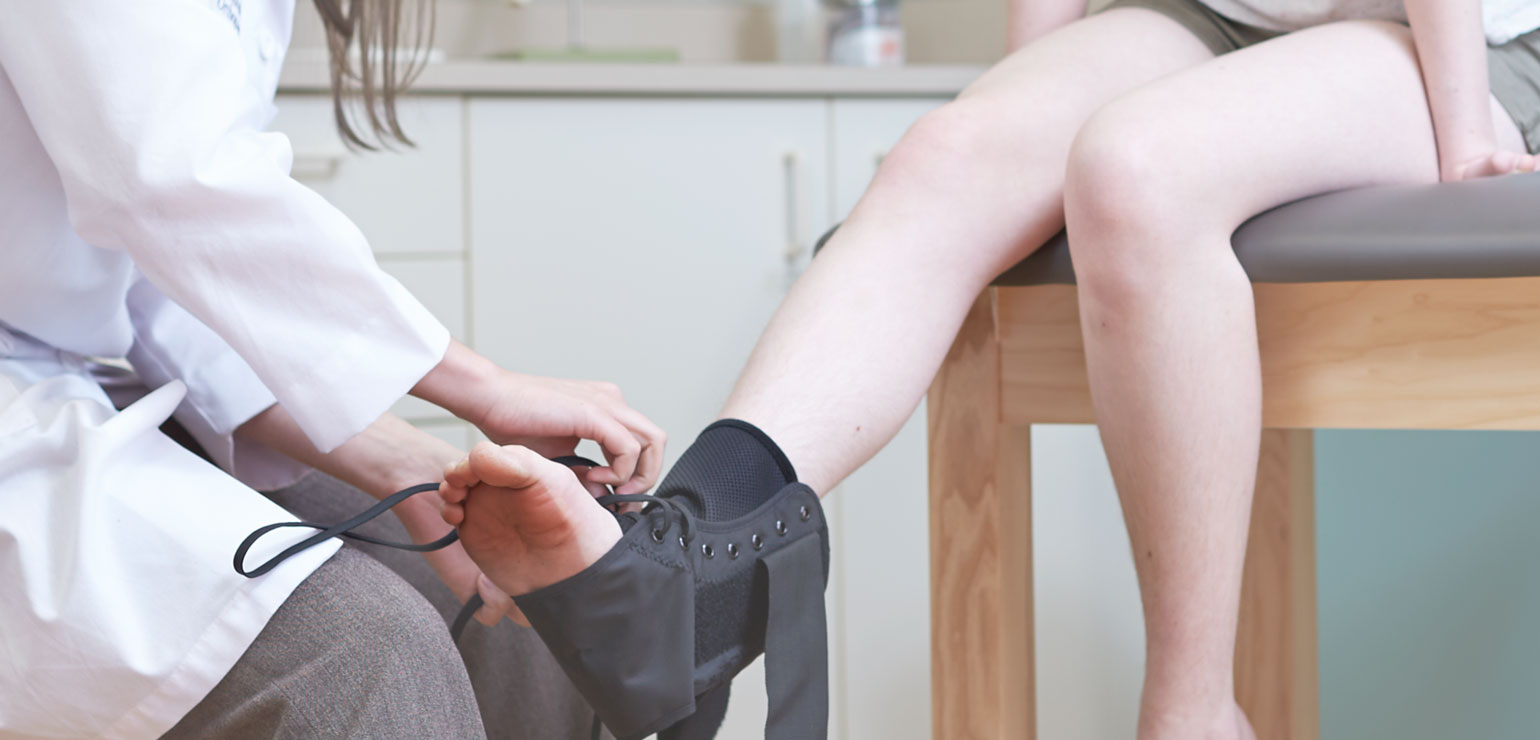
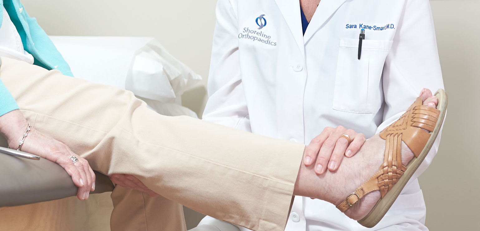
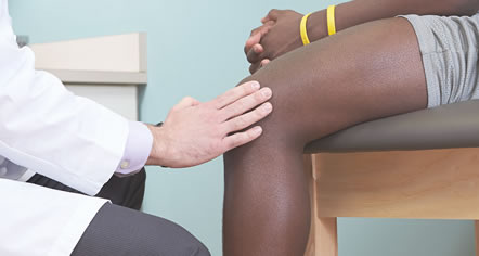
Shoreline Orthopaedics provides more comprehensive services, state-of-the-art options, technologies and techniques than anyone else in the area.
- Patient Information
- New Patient Forms
- Miscellaneous Patient Forms
- Insurance
- Patient Privacy
- Financial Policy
- How Will Weight Loss Affect Your Health
- Falls and Prevention
- Osteoporosis
- Negative Effects of Smoking
- FAQ
- Testimonials
The following information is provided to help you understand what you can expect from us regarding policies and procedures, and also what is expected of you before and after treatment or procedures.
- Education
- Protocols
- Instructional Animations
- Glossary
The following information is provided to help you gain a better understanding of anatomy, terminology, certain orthopaedic procedures, and more. If you have any questions, feel free to ask your physician.
- Protocols
- Contact
- Blog